
x ray positioning chart with images pdf
An X-ray positioning chart with images is a visual guide that illustrates optimal patient positioning for accurate radiographic imaging, ensuring diagnostic clarity and patient safety effectively always.
What is an X-ray Positioning Chart?
An X-ray positioning chart is a visual guide that outlines the optimal placement of patients for specific radiographic projections, ensuring accurate and clear imaging results while maintaining patient safety and diagnostic efficiency through precise anatomical alignment and standardized techniques.
Purpose of an X-ray Positioning Chart with Images
The purpose of an X-ray positioning chart with images is to provide radiographers with a clear, step-by-step visual reference, ensuring consistent, high-quality imaging by accurately aligning patient anatomy with imaging goals, reducing errors, and enhancing diagnostic accuracy for various radiographic projections efficiently.

Importance of Proper X-ray Positioning
Proper X-ray positioning ensures accurate image quality, enhances diagnostic accuracy, and prioritizes patient safety by minimizing radiation exposure and optimizing anatomical alignment for clear radiographic results.
Accuracy in Diagnosis
Proper X-ray positioning ensures clear, detailed images, enabling precise diagnoses. Accurate alignment of anatomical structures minimizes distortions, allowing radiologists to identify abnormalities confidently. This reduces misdiagnosis risks and unnecessary procedures, improving patient outcomes significantly.
Patient Safety
Proper X-ray positioning minimizes radiation exposure, ensuring patient safety. Correct alignment reduces the need for repeat images, lowering radiation dose. This adherence to the ALARA principle protects patients from unnecessary exposure, promoting safer diagnostic procedures and better health outcomes overall.
Diagnostic Efficiency
Accurate X-ray positioning enhances diagnostic efficiency by ensuring clear, detailed images. Proper alignment reduces retakes, saving time and resources. Clear visuals enable quick identification of abnormalities, aiding in timely diagnoses and effective treatment planning, ultimately improving patient outcomes and streamlining clinical workflows efficiently.

Key Anatomical Landmarks for X-ray Positioning
Key anatomical landmarks guide precise X-ray positioning, ensuring accurate imaging. Key structures include thoracic vertebrae, iliac crests, and femoral heads, aiding in proper alignment for clear radiographic results effectively.
Thoracic Landmarks
Thoracic landmarks include the clavicles, scapulae, and vertebral bodies. Proper alignment ensures accurate chest and spinal imaging. The right middle lobe touches the heart border, while the lingular segment aligns with the left heart border. These references guide precise X-ray positioning for clear diagnostic results, enhancing image quality and patient safety effectively always.
Abdominal Landmarks
Abdominal landmarks like the diaphragm, liver, spleen, and digestive organs guide X-ray positioning. Proper alignment of these structures ensures accurate imaging of the abdominal cavity. Landmarks help technicians position patients correctly, optimizing image quality and diagnostic accuracy while maintaining patient safety during radiographic procedures, as outlined in detailed X-ray positioning charts with visual references. Always ensure precise anatomical alignment for clear results.
Extremities Landmarks
Extremities landmarks, such as joints and long bones, are crucial for accurate X-ray positioning. The alignment of the humerus, femur, tibia, and fibula ensures proper imaging of arms and legs. Landmarks like the shoulder, elbow, knee, and ankle guide technicians to achieve clear, distortion-free images, enabling precise diagnosis of fractures or joint abnormalities while maintaining patient safety and image quality. Always refer to detailed charts for optimal positioning.
Common X-ray Projections and Their Purposes
Chest X-rays diagnose lung and heart issues, spinal X-rays assess vertebral alignment and injuries, and limb X-rays evaluate fractures and joint conditions, ensuring accurate diagnoses and targeted treatments.
Chest X-rays
Chest X-rays diagnose lung disorders like pneumonia, tuberculosis, and fractures, as well as heart conditions. Proper positioning involves the patient standing upright with hands on hips, ensuring full inspiration for clear lung visualization. The chart provides detailed visuals and guidelines to ensure accurate imaging and proper alignment, essential for diagnosing thoracic conditions effectively and safely.
Spinal X-rays
Spinal X-rays diagnose fractures, herniated discs, and scoliosis, requiring precise patient positioning. Patients may stand or lie prone/supine, ensuring the spine aligns with the X-ray beam. Charts provide visual guidance for accurate alignment, helping technicians capture clear images of vertebral structures, enhancing diagnostic accuracy while minimizing retakes and ensuring patient safety during the imaging process always.
Limbs and Joints X-rays
Limbs and joints X-rays diagnose fractures, arthritis, or injuries, requiring precise positioning. Patients may stand, sit, or lie down, with the affected area centered on the image receptor. Proper alignment ensures clear visualization of bone structures, aiding in accurate diagnoses and minimizing retakes, while maintaining patient comfort during the imaging process effectively always.
How to Read an X-ray Positioning Chart
Reading an X-ray positioning chart involves interpreting symbols, aligning anatomy with imaging goals, and using visual aids to ensure accurate patient positioning and optimal image quality.
Understanding Symbols and Markings
Understanding symbols and markings on an X-ray positioning chart is crucial for accurate imaging. These include arrows, lines, and anatomical indicators that guide correct patient alignment. Symbols denote beam direction, body part orientation, and reference points, ensuring proper positioning. They help technicians align the patient accurately with the X-ray beam for clear, diagnostic images every time.
Interpreting Patient Positioning
Interpreting patient positioning involves understanding how the body is aligned for X-ray imaging. Positions like prone, supine, erect, or lateral describe how the patient is placed. Accurate positioning ensures the X-ray beam captures the correct anatomical structures, reducing retakes and improving diagnostic clarity. Proper alignment is critical for obtaining clear, usable images while minimizing radiation exposure risks.
Aligning Anatomy with Imaging Goals
Aligning anatomy with imaging goals ensures the X-ray captures the desired structures clearly. Proper positioning guides the X-ray beam to highlight specific areas, optimizing image quality. This alignment minimizes distortion and ensures diagnostic accuracy, making it easier to identify abnormalities. Clear visuals aid healthcare providers in making precise diagnoses and treatment plans effectively every time.

X-ray Exposure Factors and Their Impact
X-ray exposure factors like kVp, mA, and exposure time significantly influence image quality; Proper optimization ensures clarity, reducing retakes and enhancing diagnostic accuracy effectively always, improving patient outcomes.
Factors Affecting Image Quality
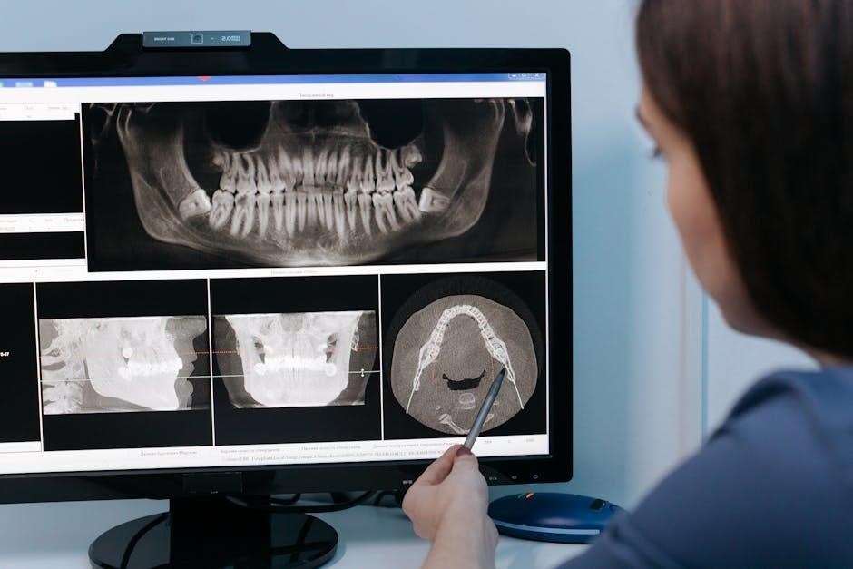
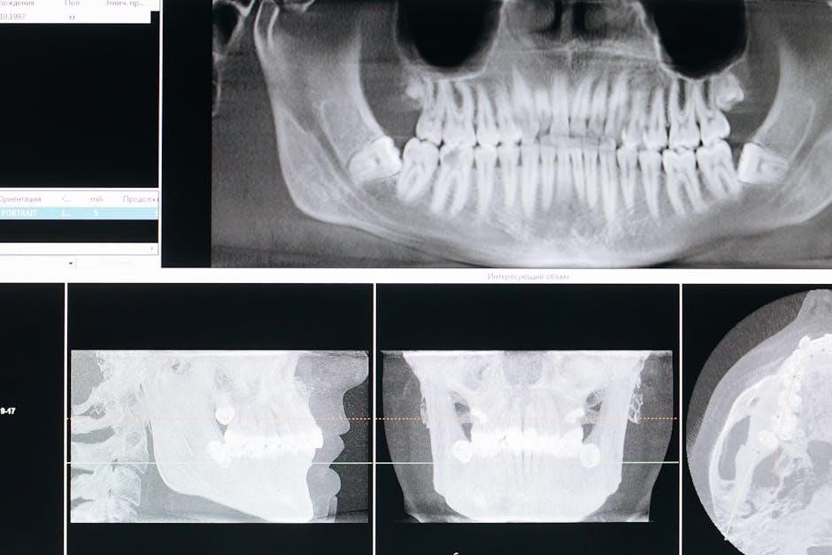
Image quality in radiography is influenced by patient movement, positioning accuracy, equipment calibration, and anatomical variations. Proper alignment ensures clear visualization, while poor positioning or equipment malfunctions can degrade image clarity. Modern systems, like the Dürr VistaScan Plus, use advanced image plate technology to optimize quality, even with challenging patient conditions, ensuring precise diagnostic results always.
These factors highlight the importance of precise techniques and patient cooperation for optimal imaging outcomes.
Exposure Techniques for Different Projections
Exposure techniques vary based on the projection type, ensuring optimal image quality. Chest X-rays require precise patient positioning, while spinal and limb projections demand adjusted angles and doses. Modern systems, like the Dürr VistaScan Plus, use image plate technology for consistent results. Proper exposure ensures diagnostic clarity, minimizing retakes and radiation exposure effectively.
Techniques must adapt to patient anatomy and projection goals for accurate imaging outcomes.

Digital vs. Conventional X-ray Systems
Digital systems offer superior image clarity, reduced radiation exposure, and faster processing compared to conventional film-based systems, enhancing diagnostic efficiency and patient safety significantly.
Advantages of Digital Systems
Digital X-ray systems provide superior image quality, reduced radiation exposure, and faster processing. They enable image manipulation and enhancement, improving diagnostic accuracy. Digital systems also allow for easier image sharing and storage, enhancing workflow efficiency. Additionally, they support environmentally friendly practices by eliminating the need for film and chemicals, making them a sustainable choice for modern radiography.
Limitations of Conventional Systems
Conventional X-ray systems rely on film and chemicals, increasing costs and environmental impact. They lack the ability to digitally manipulate images, potentially requiring retakes for optimal clarity. Additionally, conventional systems often result in higher radiation exposure and longer processing times, delaying diagnosis and reducing workflow efficiency compared to digital alternatives.
X-ray Positioning Devices Market Trends
The market is growing due to increasing demand for precise imaging and technological advancements, with expectations of robust growth driven by improved diagnostic accuracy and patient safety.
Current Market Overview
The X-ray positioning devices market is experiencing steady growth, driven by advancements in digital imaging and increasing demand for precise patient positioning. Innovations like the Dürr VistaScan Plus and integration of AI are shaping the industry, with a focus on improving diagnostic accuracy and operational efficiency across healthcare facilities globally.
Future Growth Predictions
The X-ray positioning devices market is projected to expand significantly, fueled by technological advancements in digital radiography and AI integration. Increased adoption of 3D imaging and personalized positioning solutions will drive growth, with a focus on enhancing patient outcomes and streamlining radiographic workflows in the coming years.

Common Mistakes in X-ray Positioning
Patient misalignment and insufficient exposure are frequent errors, leading to poor image quality and potential misdiagnosis. Proper training and adherence to positioning charts are essential to minimize these issues.
Patient Misalignment
Patient misalignment occurs when the body is not positioned correctly, leading to poor image quality and potential misdiagnosis. This common mistake can result in inaccurate diagnostic results. Proper training and adherence to positioning charts are essential to minimize such errors and ensure accurate radiographic outcomes.
Insufficient Exposure
Insufficient exposure refers to inadequate X-ray penetration, resulting in underexposed images; This can obscure anatomical details, making diagnosis difficult. Factors like incorrect kVp or mAs settings, improper patient positioning, or equipment malfunctions may cause this. Proper calibration and adherence to exposure charts are crucial to achieve optimal image quality and accurate diagnostic results.

Radiation Safety in X-ray Positioning
Radiation safety in X-ray positioning involves minimizing exposure to ionizing radiation through proper techniques, protective gear, and adherence to ALARA principles to ensure patient and technician safety.
ALARA Principle
The ALARA principle emphasizes minimizing radiation exposure to patients and technicians during X-ray positioning. It involves optimizing techniques, using protective gear, and adjusting exposure factors to achieve diagnostic image quality while keeping doses as low as reasonably achievable, ensuring safety without compromising accuracy or efficiency in radiographic procedures.
Patient and Technician Protection
Patient and technician protection in X-ray positioning involves using lead aprons, thyroid collars, and gloves to shield against radiation. Proper positioning minimizes exposure, while digital systems reduce doses. Regular equipment maintenance and adherence to safety protocols further ensure a secure environment, balancing diagnostic needs with radiation safety for all involved in the imaging process.
Clinical Applications of X-ray Positioning Charts
Clinical applications of X-ray positioning charts include emergency radiography, orthopedic imaging, and routine diagnostics, ensuring accurate and efficient patient assessment across various medical specialties and scenarios effectively.
Emergency Radiography
X-ray positioning charts are crucial in emergency radiography, providing rapid, accurate patient positioning guides for acute injuries or conditions. They ensure clear imaging of chest, abdominal, and extremity trauma, enabling swift diagnosis and treatment. These charts help reduce errors and improve outcomes in high-stress environments, making them indispensable in emergency care settings worldwide every day.
Orthopedic Imaging
X-ray positioning charts are essential in orthopedic imaging, ensuring precise alignment of bones and joints. They guide optimal angles for capturing detailed images of fractures, degenerative conditions, and joint alignments; Accurate positioning enhances diagnostic clarity, aiding orthopedic specialists in developing effective, personalized treatment plans tailored to specific patient needs and conditions effectively.
How to Create an Effective X-ray Positioning Chart
Designing an effective X-ray positioning chart involves clear visual aids, precise anatomical references, and standardized protocols. Prioritize simplicity, using symbols and images for clarity. Tailor the chart to specific equipment and update regularly to reflect new techniques. Ensure patient safety guidelines are emphasized, and collaborate with experts for accuracy and relevance, ensuring it remains a practical tool for radiographers.
Design Principles
Effective X-ray positioning charts emphasize simplicity and clarity. Use high-quality images, clear symbols, and concise labels. Organize content logically, ensuring readability and ease of use. Incorporate color coding for differentiation and highlight key anatomical landmarks. Ensure consistency in terminology and formatting. Adhere to standardized positioning protocols and update regularly to reflect advancements in radiography. Prioritize patient safety and diagnostic accuracy.
Incorporating Visual Aids
Visual aids like high-quality images, diagrams, and symbols enhance the clarity of X-ray positioning charts. Use arrows and labels to indicate precise patient positioning and alignment. Incorporate anatomical landmarks and reference lines for accuracy. Ensure images are clear and well-lit, with adaptable views for various projections. This approach simplifies comprehension and improves the chart’s practical application for radiographers.
Updating and Refining the Chart
Regularly update the X-ray positioning chart with new imaging techniques and technologies. Incorporate feedback from radiographers and physicians to refine accuracy. Review and revise anatomical landmarks and projections to reflect current standards. Ensure all updates align with patient safety and diagnostic efficiency, maintaining the chart as a reliable, evolving resource for precise imaging practices always.

Factors Affecting Image Quality
Patient positioning, exposure settings, and equipment quality significantly impact image clarity. Proper calibration, skilled operation, and patient cooperation are crucial for accurate and diagnostic images.
Patient Cooperation
Patient cooperation is crucial for achieving clear X-ray images. Proper positioning, remaining still, and following instructions ensure accurate results. Cooperation reduces retakes and enhances diagnostic quality, improving patient safety and efficiency in imaging procedures.
Equipment Calibration
Proper calibration of X-ray equipment ensures optimal image quality and patient safety. Digital systems with image plate technology require precise adjustments to deliver accurate results. Regular maintenance and adherence to manufacturer guidelines are essential for consistent performance, minimizing errors, and reducing radiation exposure during imaging procedures.
Future Directions in X-ray Positioning
Future advancements include AI-driven positioning systems and 3D imaging integration, enhancing accuracy and reducing radiation exposure while improving diagnostic outcomes and patient care significantly.
AI in Positioning
AI enhances X-ray positioning by using machine learning algorithms to analyze patient anatomy, predict optimal angles, and adjust settings, reducing retakes and improving image quality. AI systems can integrate with digital X-ray devices, ensuring precise alignment and minimizing radiation exposure. This technology streamlines workflows, improves diagnostic accuracy, and enhances patient safety, making radiography more efficient and reliable overall.
3D Imaging Integration
3D imaging integration enhances X-ray positioning charts by providing detailed volumetric views, improving accuracy in complex anatomical assessments. This technology complements traditional 2D imaging, allowing better visualization of structures like joints and fractures. Advanced systems, such as the Dürr VistaScan Plus, offer panoramic and cephalometric capabilities, reducing retakes and improving diagnostic confidence for precise patient care and treatment planning.
Proper use of X-ray positioning charts ensures accurate, safe, and efficient radiographic imaging, supported by advancements like 3D integration, enhancing diagnostic confidence and patient care outcomes significantly.
An X-ray positioning chart with images serves as a visual guide, ensuring precise patient alignment for accurate radiographic imaging. It highlights key anatomical landmarks, standardizes positioning techniques, and covers various projections, from chest to spinal X-rays. By optimizing diagnostic accuracy and safety, these charts are essential for radiographers, enhancing imaging quality while minimizing radiation exposure effectively.
Future Implications
Future advancements in X-ray positioning charts will likely integrate AI for automated adjustments and 3D imaging for enhanced accuracy. These innovations promise personalized patient positioning, reducing radiation exposure and improving diagnostic outcomes. As technology evolves, digital systems will streamline workflows, offering real-time feedback and higher precision in radiographic imaging, revolutionizing healthcare diagnostics and patient care significantly.